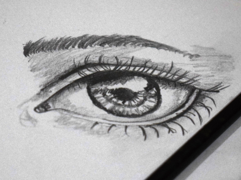
The sagittal vertical (height) of a human adult eye is approximately 23.7 mm (0.93 in), the transverse horizontal diameter (width) is 24.2 mm (0.95 in) and the axial anteroposterior size (depth) averages 22.0–24.8 mm (0.87–0.98 in) with no significant difference between sexes and age groups. The eyeball is generally less tall than it is wide. The size of the eye differs among adults by only one or 2 millimetres. Photons of light falling on the light-sensitive cells of the retina ( photoreceptor cones and rods) are converted into electrical signals that are transmitted to the brain by the optic nerve and interpreted as sight and vision. The lens shape is changed for near focus (accommodation) and is controlled by the ciliary muscle. Light energy enters the eye through the cornea, through the pupil and then through the lens. The size of the pupil, which controls the amount of light entering the eye, is adjusted by the iris' dilator and sphincter muscles. The iris is the pigmented circular structure concentrically surrounding the centre of the eye, the pupil, which appears to be black. An area termed the limbus connects the cornea and sclera. The posterior chamber constitutes the remaining five-sixths its diameter is typically about 24 mm (0.94 in). The cornea is typically about 11.5 mm (0.45 in) in diameter, and 0.5 mm (500 μm) in thickness near its centre. The cornea is transparent and more curved and is linked to the larger posterior segment, composed of the vitreous, retina, choroid and the outer white shell called the sclera. The anterior segment is made up of the cornea, iris and lens.

The eye is not shaped like a perfect sphere, rather it is a fused two-piece unit, composed of an anterior (front) segment and the posterior (back) segment. The front part is also called the anterior segment of the eye. A thin layer called the conjunctiva sits on top of this. The front visible part of the eye is made up of the whitish sclera, a coloured iris, and the pupil. There are six extraocular muscles that control eye movements. The eyes sit in bony cavities called the orbits, in the skull. Humans have two eyes, situated on the left and the right of the face.
#Eye sketch full#
Three types of cells in the retina convert light energy into electrical energy used by the nervous system: rods respond to low intensity light and contribute to perception of low-resolution, black-and-white images cones respond to high intensity light and contribute to perception of high-resolution, coloured images and the recently discovered photosensitive ganglion cells respond to a full range of light intensities and contribute to adjusting the amount of light reaching the retina, to regulating and suppressing the hormone melatonin, and to entraining circadian rhythm. The remaining components of the eye keep it in its required shape, nourish and maintain it, and protect it. The retina makes a connection to the brain via the optic nerve. In order, along the optic axis, the optical components consist of a first lens (the cornea-the clear part of the eye) that accomplishes most of the focussing of light from the outside world then an aperture (the pupil) in a diaphragm (the iris-the coloured part of the eye) that controls the amount of light entering the interior of the eye then another lens (the crystalline lens) that accomplishes the remaining focussing of light into images then a light-sensitive part of the eye (the retina) where the images fall and are processed.

It is approximately spherical in shape, with its outer layers, such as the outermost, white part of the eye (the sclera) and one of its inner layers (the pigmented choroid) keeping the eye essentially light tight except on the eye's optic axis. The eye can be considered as a living optical device. The human eye is a sensory organ, part of the sensory nervous system, that reacts to visible light and allows humans to use visual information for various purposes including seeing things, keeping balance, and maintaining circadian rhythm.


 0 kommentar(er)
